


Electrical Impedance Tomography (EIT)
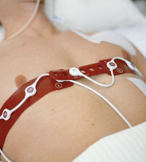
Fig. 1:Picture from [EML15]
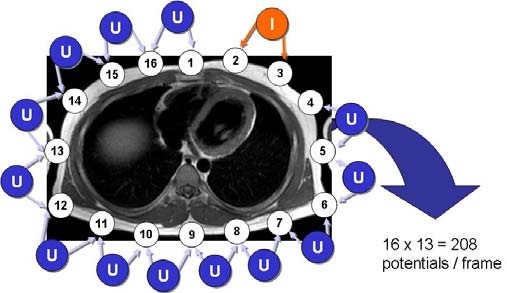
Fig. 2: Picture from [Leo+12]

Fig. 3: Picture from [LL12]
Contact persons
Project description
In critically ill patients, a continuous monitoring is desirable. This is especially true in mechanically ventilated patients in the intensive care unit (ICU). Currently, ventilation parameter such as ventilation pressure and volume are adjusted using global parameters such as the results of blood gas analysis and the lung compliance. In order to improve the patients outcome, not only the ventilated volume, but also the distribution of the volume inside the lung should be monitored. This is especially important in patients suffering from lung diseases such as pneumonia, lung edema and atelectasis. Since EIT is non-invasive and can be used at bedside, it is a promising imaging technique in the ICU.
Methods
Electrical Impedance Tomography (EIT) is a non-invasive imaging method that allows to monitor changes of electrical impedance within a cross-section spanned by a set of electrodes (typically n = 16 or 32) attached around the human torso. Earlier, single electrodes with individual cables were placed on the patient’s skin. Modern EIT devices integrate multiple electrodes into a special belt, which allows quicker handling and more accurate measurements, since electrodes are placed equidistantly in a reproducible way.
As it is shown in fig.1, the belt is usually placed at the level of the 4th to 6th intercostal space (”anatomic space between two ribs”) and therefore includes large areas of the lungs as well as the heart in the imaged region. One additional reference electrode is placed in greater distance to the belt and makes sure that all voltage measurements are made with respect to the same ground potential. Contact gel can be used to improve the electrical connection between electrode surface and the patient’s skin.
The EIT measurement principle is illustrated in fig.2. A so-called ”adjacent drive configuration” is used. A small alternating current (I) is injected (”driven”) into two neighbouring (”adjacent”) electrodes (here 2 and 3) and differential voltages (U) are measured at all remaining 13 electrode pairs. After all 13 voltages are measured, the current injection is shifted on to the next electrode pair (here 3 and 4) and voltage measurements at the remaining electrodes are repeated. Current injection is shifted circularly until all 16 electrode pairs were used for current injection once. For each current injection, voltage measurements at the remaining 13 electrode pairs are performed. This results in a set of 16 × 13 = 208 voltage measurements, which is called a frame.
Reconstruction
In principle, two problems can be addressed in EIT. With the knowledge of the conductivity distribution inside the body and the injected current, the resulting measured EIT voltages can be calculated. This is called the forward problem and describes the calculation of voltages on the body surface from a known conductivity distribution. The forward problem is solvable, for example by using the finite element method. In EIT, however, we are interested in reconstructing the conductivity distribution from the obtained voltage measurements. This is the so-called inverse problem. Unfortunately, this problem is ill-posed and not solvable analytically. The solving of the inverse problem is the heart of the reconstruction. A reconstruction example of a human thorax and a pig torso is shown in fig.3.
Further Literature
- [Leo+12] Steffen Leonhardt et al. “Electrical Impedance Tomography for Hemodynamic Monitoring”. In: 2012 Annual International Conference of the IEEE Engineering in Medicine and Biology Society. Vol. 2012. IEEE, Aug. 2012, pp. 122–125. isbn: 978-1-4577-1787-1. doi: 10.1109/EMBC.2012.6345886.
- [LL12]; Leonhardt, S., & Lachmann, B. (2012). Electrical impedance tomography: The holy grail of ventilation and perfusion monitoring? Intensive Care Medicine, 38(12), 1917–1929. doi.org/10.1007/s00134-012-2684-z
- [EML15] Eckhard Teschner, Imhoff Michael, and Steffen Leonhardt. Electrical Impedance Tomography: The Realisation of Regional Ventilation Monitoring (2nd Edition). Tech. rep. Draeger GmbH, 2015.